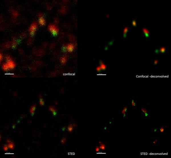
Stimulated Emission Depletion (STED) microscopic images can now be deconvolved with the new Huygens STED option with truly stunning results. You can find the latest version at the Download page under development versions.
Developed bij Prof. Stefan Hell and now manufactured by Leica Microsystems, STED microscopes offer true super-resolution. The unique feature of the STED microscope is that it does not have a bandwidth limit, i.e. a barrier beyond which object details are not imaged anymore. While other super-resolution systems are still hampered by this wavelength dependent limit, a STED microscope moves happily beyond it.
Leica now offers two types of STED microscopes of which the CW version allows the usage of all kind of fluophores in the green range thus enabling full biological capabilities.
The Z-resolution, a very important part of biological research, is currently on the level of confocal Z-resolution. By applying Huygens STED deconvolution option you will get a huge increase in contrast (~10 times), more resolution in Z, and also improve the already high lateral STED resolution.
As always, Huygens deconvolution reduces noise and blurring, and takes depth-dependent spherical aberration into account. Geometrical distortions can also be easily corrected in Huygens.

You are welcome to explore the new Huygens STED option using a version.
Developed bij Prof. Stefan Hell and now manufactured by Leica Microsystems, STED microscopes offer true super-resolution. The unique feature of the STED microscope is that it does not have a bandwidth limit, i.e. a barrier beyond which object details are not imaged anymore. While other super-resolution systems are still hampered by this wavelength dependent limit, a STED microscope moves happily beyond it.
Leica now offers two types of STED microscopes of which the CW version allows the usage of all kind of fluophores in the green range thus enabling full biological capabilities.
The Z-resolution, a very important part of biological research, is currently on the level of confocal Z-resolution. By applying Huygens STED deconvolution option you will get a huge increase in contrast (~10 times), more resolution in Z, and also improve the already high lateral STED resolution.
As always, Huygens deconvolution reduces noise and blurring, and takes depth-dependent spherical aberration into account. Geometrical distortions can also be easily corrected in Huygens.
Huygens Confocal and STED deconvolution
Example images obtained from Dr. Juraj Kabat (Biological Imaging Facility, NIH/NIAID, Bethesda, USA), who tested the Huygens STED deconvolution option and commented on the results: "I am really excited" and "it is clearly visible that the STED image is giving much more spatial details, although they are visible only after deconvolution".Comparision of confocal and STED images, before and after deconvolution with Huygens.
Signal shows regulatory proteins Dmc1 and Rad51 for DNA repair during homologous chromosome recombination in meiosis. Dmc1 shown in green: Mega 520 - 532/685 for ex/em and 770 for depletion. Rad51 shown in red: Atto 647 640/685 ex/em and 770 for depletion. Courtesy of Dr. Juraj Kabat (Biological Imaging Facility, NIH/NIAID, Bethesda, USA.)
You are welcome to explore the new Huygens STED option using a
Permalink: https://svi.nl/blogpost28-STED-deconvolution-now-available-in-Huygens