4Pi microscope
A 4Pi microscope is one type of Fluorescence Microscope in which the principle of a Confocal Microscope is applied trying to approach the ideal case of spherical wavefronts.
"In this method, two high-resolution objectives are positioned across from each other, but directed on the same spot in such a way that an interference occurs in their joint focus. This allowed them to increase the resolution of Fluorescence Microscopes by a factor of 3 to 7 along the optical axis (Z) and thus to generate correspondingly thinner slices in a 3D image stack" 1

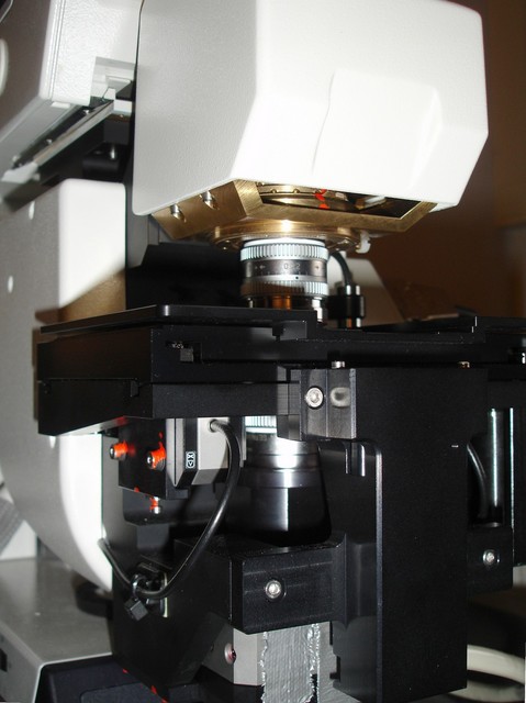
The Leica 4Pi microscope at the Optical Imaging Centre at Erasmus Medical Center. Courtesy Dr. Gert van Cappellen.
"The idea behind our 4Pi-microscope is to mimic the 'close to ideal' situation by using two opposing objective lenses coherently, so that the two wavefronts add up and join forces ... Allowing the illumination wavefronts to constructively interfere in the sample produces a main focal spot that is sharper in the z-direction by about 3-4 times (4Pi of type A). A similar improvement is obtained if the lenses add their collected fluorescence wavefronts in a common point detector (4Pi of type B). Doing both together is best, of course, and leads to a 5-7-fold improvement of resolution along z (4Pi of type C)". 2
For Point Spread Functions of this microscope, see Four Pi Psf.
1 http://www.uchsc.edu/lightmicroscopy/sted4pi.htm 2 http://www.mpibpc.gwdg.de/abteilungen/200/4Pi.htm
