
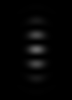
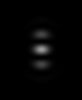
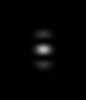
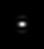
4pi PSF
Calculated Point Spread Functions for a Four Pi Microscope changing the Numerical Aperture.
"If the two lens segments were full spherical halves, the focal spot would be a (nearly) spherical spot, too. But since a considerable solid angle is not provided by the lenses, interference typically spawns off 2 axial side-lobes which, if not taken into account, lead to artefactual images" (1).
All images are 1 µm height. α is the angular aperture of the 4pi lens (the total angle of the two segments together as measured from the focus).
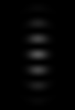
 α = 194 ° </td><td> α = 212 ° </td><td> α = 252 ° </td><td> α = 288 ° </td><td> α = 352 °
This is a PSF (with some intensity decay in the laterals of the convergent lens due to energy redistribution), not a theoretical 4pi illumination (where intensity would be uniform from all directions of space), but still suggests the real possibility of exciting a central point without noticeable lateral sidelobes.
Sample Size: 10 x 10 x 10 nm3 Pinhole Radius: 100 nm Lens Refractive Index: 2 Medium Refractive Index: 2 Excitation Wavelength: 488 Emission Wavelength: 520 Excitation Photons: 1 Numerical Aperture = Lens Refractive Index * sin(α/4)
1. For more information see https://nanobiophotonics.mpibpc.mpg.de/popular-reading