
Image enhancement versus deconvolution
Image enhancement is different from Image Restoration in that it is designed to emphasize features of the image that make the image more pleasing to the observer, but not necessarily to produce realistic data from a scientific point of view. Image enhancement techniques (like contrast stretching or de-blurring by a nearest neighbor procedure) provided by "Imaging packages" use no a priori model of the process that created the image.
On the other hand, techniques as DeConvolution try to model the Image Formation process and to relocate light intensities in a realistic way. See the following comparison:
"Estimating the underlying fluorescence signal: Figure 1(a) is a typical image of the optic disc in the retina of a transgenic mouse where the fluorescence signal is from the GFP protein which, in this case, labels the glial cells. As observed, the image has a very low SNR and this makes it extremely difficult to detect any suitable landmark points and it is clearly inappropriate to use such images as reference images in rank matching. An a-priori estimate is therefore required to accurately estimate the underlying fluorescence signal. We have explored several methods of determining this estimate and found that the Huygens deconvolution method (Huygens Essential Software, Scientific Volume Imaging BV) gives the best results."
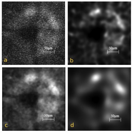 Fig. 1. (a) Input image, of the GFP signal from the optic disc of the transgenic mouse. (b)-(d) restored image obtained after subjecting the input image to (b) Huygens deconvolution (c) 9 × 9 median filtering and (d) speckle reducing anisotropic diffusion.
"The deconvolved result of Fig. 1(a) is shown in Fig. 1(b) and as observed, the speckle noise is removed and the fluorescence intensity profile is characterized by various intensity maxima. Conversely, a 9 × 9 median filtering method of estimating the a-priori estimate fails to remove the speckle noise and instead distorts the image as observed in Fig. 1(c). Figure 1(d) shows the results of applying anisotropic diffusion where the noise appears to be removed but at the expense of diffusing the underlying fluorescence signal resulting in the image appearing blurred overall." (S. Kumar, G. Ho, K.M. Woo and L. Zhuo, Institute of Bioengineering and Nanotechnology, Singapore, Optics Express, Vol. 16, Issue 11, pp. 8250-8262).