SVI Screenshot Gallery
| Screenshot | Description window |
|---|---|
| The Huygens Essential main window. Huygens Essential combines deconvolution with a graphical wizard driven user interface for parameter editing and deconvolution. Thumbnail representations of the original image and deconvolved result are placed in the grey work area in the left part of the main window, and tabs on the right show various image parameters. Visualization and Analysis tasks are done in separate windows for each image. |
|
| The Huygens Professional main window. Huygens Professional combines a graphical user interface (GUI) designed to provide you rapid access to microscopic image restoration, and additional processing and analysis tasks with the extreme flexibility of the Tool Command Language (Tcl) through a versatile Tcl Shell. The GUI shows all images present in a compact scrollable window. Just by selecting one or more of these you can accomplish most of the simple housekeeping operations like copying images or splitting multi-parameter images that go with image processing. A Task Report pane shows the output of some commands, and the intelligent Tcl Shell lets you type Tcl Huygens commands to quickly operate with all images. |
|
| The SVI support Wiki pages. The SVI-wiki is a rapidly expanding public knowledge resource on 3D microscopy, image restoration (deconvolution), visualization and analysis. Based on the wiki principle, it is open to contributions from every visitor. The SVI support Wiki pages can be found here: http://www.svi.nl/FrontPage |
|
| Measuring a line profile in the Twin Slicer. The Twin Slicer allows you to synchronize views of two images, measure distances, plot line profiles, etc. In Basic Mode, image comparison is intuitive and easy, while the Advanced Mode gives the user the freedom to rotate the cutting plane to any arbitrary orientation, link (synchronize) or unlink viewing parameters between the two images, and more. |
|
| Twin Slicer in overview mode. An easy 'overview' mode can be configured as follows: - Select the same image for both the left and right slicer. - Tick 'Advanced mode' and tick all links in the 'View' menu, except for 'Zoom' and 'Panning'. - Drag the sash to the left to make the left slicer a bit smaller. - Click the 'Zoom' tab at the bottom and click the 'Zoom all' button. Now you can use the right slicer to zoom in on your data, while the left slicer shows where you are. |
|
| Orthogonal Slicer. The Orthogonal Slicer is designed to show the same point in 3D space from 3 orthogonal directions (axial; top left, frontal; bottom left, and transverse; bottom right). If you move one of the slices, the others will follow to make sure that the center of each of the slices intersects in the same point in space. This behaviour makes the Ortho Slicer a useful tool to study small objects in 3D. |
|
| The Huygens Movie Maker. The Movie Maker is a tool that allows you to easily create sophisticated animations of your multi-channel 3D images using the powerful SFP, MIP, and surface renderers. The AVI movies can be played by Windows Media Player, Apple Quicktime, and other players that support the MJPEG standard. |
|
| MIP Renderer. The maximum intensity projection (MIP) renderer projects the voxels with maximum intensity that fall in the way of parallel rays traced from the viewpoint to the plane of projection. This allows you to obtain a direct spatial projection of your 3D microscopy data from the pointview you wish. |
|
| SFP Renderer. This renderer is based on taking the 3D microscopy image as a distribution of fluorescent material, simulating what happens if the material is excited and how the subsequently emitted light travels to the observer. The computational work is done by the Simulated Fluorescence Process (SFP) algorithm. The unique properties of this algorithm enable it to create depth cue rich images from unprocessed data. Because it does not rely on boundaries or sharp gradients, it is eminently suited to render 3D microscopic data sets. Since the SFP algorithm is bases on ray-tracing it does not require a special graphical board as the polygon based techniques do. |
|
| Surface Renderer. The Surface Renderer enables you to represent your microscopy data in a convenient way to clearly see separated volumes. It is not only capable of iso-surface rendering; it is also able to show MIP projections together with the surfaces to be used as a reference to the original microscopic voxel data. Because the Surface Renderer is based on rendering continuous surfaces with fast ray tracing algorithms, there is no need for any special graphic card. The fast ray tracers can utilize 64 bit multiprocessor systems, and are therefore able to render very large microscopic volume data to high resolution output images. |
|
| Batch Processor. The Huygens Batch Processor automates the running of multiple deconvolution tasks. A small wizard guides you in creating tasks. Once the Task List is complete pressing 'Run' will schedule the tasks for processing immediately or at a later moment as determined by the timer. If you have multiple processors in your computer the batch processor can run several tasks simultaneously or sequentially. While the queue processed it is still possible to delete, stop or add new tasks. |
|
| PSF Distiller. The Huygens PSF Distiller allows you to obtain a Point Spread Function (PSF) for your own microscope based on 3D datasets with recorded bead images. A wizard guides you in selecting your bead images creating and saving the PSF for further use in deconvolution runs. The so-called measured (or experimental) PSF allows you to improve your restoration results. The color shift is reported which can be used be used to improve colocalization studies. |
|
| Colocalization Analyzer. Colocalization analysis allows you to obtain quantitative information about the amount of spatial overlap between structures in different data channels, for 3D images and 3D-time series. As this overlap can be defined in many ways, the Colocalization Analyzer gives you the colocalization coefficients most commonly used in literature. |
|
| Object analyzer. The interactive Object Analyzer tool for 3D microscopy images in Huygens Essential and Huygens Professional allows you to obtain statistics of individual objects by clicking on them, or analyzing all objects with a single button press. |
|
| Chromatic Shift Corrector. The Chromatic Shift Corrector can estimate and correct for chromatic shifts, removing the existing misalignments across different channels. This tool reports the existing chromatic shifts as vectors. A shift vector edit tool allows a customized correction. Furthermore, the Chromatic Shift Corrector is equipped with support for templates to save a set of shift vectors or load a set of predetermined shift vectors. |
|
| CrossTalk Corrector. The CrossTalk Corrector can estimate and correct for crosstalk (i.e., bleedthrough or crossover), which is the recording of some signal as a certain dye when it really comes from a different one. This Corrector estimates the cross talk when pressing the "Estimate" button. The preview windows show the corrected 2D histogram and MIP according to the position of the two slopes in the main 2D histogram. Upon pressing the "Correct image" button a crosstalk-corrected image is generated. |
|
| Operations window. The operations window allows you to access the more complex functions in Huygens Professional. Because all operations relate to an image, this image is shown in the operations window. To the right of the image is the dialog field where you can enter function specific parameters. The values and choices displayed in the entry fields are initially set to sensible factory defaults. |
|
| The Huygens Scripting window. Huygens Scripting provides easy image processing programming with or without a user interface. Running this program with the interface is ideal for programming on desktop machines, and will allow you to benefit from the versatile Tcl Shell. By leaving out the interface you have a powerful deconvolution tool that you can easiliy run on remote machines. |
|
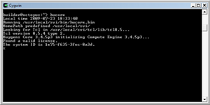
| Huygens Core. Huygens Core, part of the Huygens Software, is a full compute engine intended to run image processing and deconvolution jobs on large 64 bit multiprocessor servers without a specific graphical interface. It can be easily integrated in web applications for remote image processing, such as the Huygens Remote Manager (HRM). |
|
| HRM: a web interface for Huygens Core. Made by Huygens users, the Huygens Remote Manager is a powerful web interface for Huygens Core. With it, multiple users can benefit from the same computational resources and review their deconvolutions on-line. |

