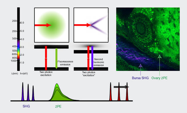
Second harmonic generation
Second harmonic generation (SHG), also called "frequency doubling", is a nonlinear optical process, in which photons interacting with a nonlinear material are effectively "combined" to form new photons with twice the energy, and therefore twice the frequency and half the wavelength of the initial photons (see also ref. 1).
SHG microscopes are Non Linear systems due to the coherent nature of the emitted light. Because of this coherence, signals arriving from different points may interfere with each another. This interfence hampers deconvolution, so one must be very careful with the interpretation of the results if nonlinearity is present and a linear convolution is assumed in the Image Formation. In principle, DeConvolution is meant for linear optical systems, however good results with Huygens have been obtained with coherent images from brighfield and SHG (see this SHG image of 1st prize winner of the 2017 Image Contest).
Comparison of SHG with Multi Photon Microscope
From 2PE fluorescence vs. second harmonic generation (SHG)

In harmonic generation, multiple photons interact simultaneously with a molecule with no absorption events. Because n-photon harmonic generation is essentially a scattering process, the emitted wavelength is exactly 1/n times the incoming fundamental wavelength. When the excitation color is changed, the emission color changes also. In contrast the wavelength of fluorescence emission is Stokes-shifted to a longer wavelength; the line shape is determined strictly by the molecular energy levels. Practically it is relatively easy to design multicolor MPM by tuning the harmonic generation so that it is well separated from the fluorescence (see mouse ovary image above — acquired in collaboration with Alexander Nikitin, Biomedical Sciences, Cornell).
Another major difference is that for scattering microscopies, the emitted light is coherent and therefore must satisfy phase matching constraints. For example, the SHG profile in MPM retains phase information and emits with a directionality dependent on the nature of the scatterers (Moreaux, L., Sandre, O. & Mertz, J. (2000) J Opt Soc Am B 17, 1685-1694. Mertz, J. & Moreaux, L. (2001) Opt Commun 196, 325-330.). Fluorescence in general emits isotropically.
Physical details
From Frequency doubling, Encyclopedia of Laser Physics and Technology
Definition: the phenomenon that an input wave can generate a wave with twice the optical frequency
Crystal materials lacking inversion symmetry can exhibit a so-called `7;(2) nonlinearity. This can give rise to the phenomenon of frequency doubling, where an input (pump) wave generates another wave with twice the optical frequency (i.e., half the wavelength) in the medium. This process is also called second-harmonic generation.
For low pump intensities, the second-harmonic conversion efficiency is small and grows linearly with increasing pump intensity, so that the intensity of the second-harmonic (frequency-doubled) wave grows with the square of the pump intensity:
$$ P_2 = \gamma P_1^2 $$
Frequency doubling is a phase-sensitive process which usually requires phase matching to be efficient. With proper phase matching and a pump beam with high intensity, high beam quality, and moderate optical bandwidth, achievable power conversion efficiencies often exceed 50%, in extreme cases even 80% (Ref. Paschotta 1994).
Read more.
Application to microscopy
From A two-photon and second-harmonic microscope, Volodymyr Nikolenko, Boaz Nemet and Rafael Yuste. Dept. of Biological Sciences, Columbia University, New York, NY 10027.
... The Olympus Fluoview/BX50WI microscope can also be modified with a minimum amount of effort to have the capability to acquire images of SHG, either from special chromophores (11, 19) (20) et al 2001, Kobayashi et al 2002) or from endogenous structures in biological tissue such as oriented collagen fibers (21). SHG, which like two-photon fluorescence is a nonlinear optical effect, is gaining recognition as an important mode of microscopy that allows biological researchers to probe a cell s trans-membrane potential (10) and thus monitor the electrical activity of nerve cells (22, 23). Unlike fluorescence, in which emitted photons are best detected with epi-illumination, SHG photons, which result from coherent scattering, are best detected in the transmission path of the microscope. One might think of the process, in simple terms, as the partial conversion of stimulating light (the IR beam) into an electromagnetic wave at twice the incident frequency (half the wavelength) with a similar bandwidth (actually times sqrt(2) ).
The SHG photons, generated at the focal spot of the laser in the sample, are collected by the condenser lens, which has to be of equal or greater numerical aperture (NA) than the objective lens NA in order to collect the whole cone of light. This is important since the SHG radiation in the forward direction (towards the condenser) is restricted to certain off-axis angles (20). We used the Olympus Aplanat Achromat oil immersion condenser with a variable NA of up to 1.4 (one does not have to use oil for NA values of less than 1). A PMT (Hamamatsu HC125-05) with the appropriate (blue) filter replaced the diffuser and fiber bundle in the auxiliary port (see schematic Figures 1b and 2). This port was originally designed for DIC imaging in transmission mode in the FLUOVIEW confocal scanner.
