
Huygens deconvolution on small beads confocal images
This is a demonstration of the capabilities of SVI's Huygens Software: we deconvolve blurry images obtained with a scanning pinhole array microscope in order to increase the resolution of the image.
Image description
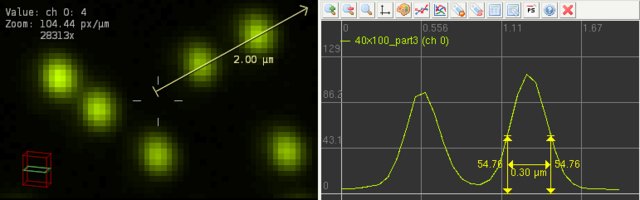
The original image was obtained with a 100x 1.4 NA lens, using an scanning array of pinholes with a diameter of 40 µm. It was sampled at 64.5 nm x 64.5 nm XY pixel size, and with a separation of 100 nm between planes in the 3D stack.
It shows a dense collection of fluorescent beads of 170 nm in diameter. Notice that with this sampling resolution, one bead should have a width of less than three pixels in its central XY plane.
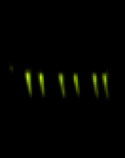
Still , due to the intrinsic blur of the Image Formation, the beads look larger than that: each bead spans over more than five pixels in XY, and the Half Intensity Width is 300 nm, as shown in this intensity profile along a central slice of the stack:
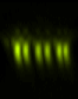
In these restoration tests we cropped a small portion of the image to show in detail a closely packed group of beads. The images below are top and lateral Maximum Intensity Projections of the 3D images. They clearly show that deconvolution improves the Spatial Resolution of the image, and restores the correct size of the beads.
Restoration results
We compare the original image with two deconvolved images:
- One using a Theoretical Psf based on the image parameters, that can reduce blur but not all other aberrations in the microscope
- Another one using an Experimental Psf distilled from the beads themselves, that can also partially restore the tilting aberration.
Despite that, as explained at Deconvolving Beads, the perfect 1:1:1 aspect ratio of a sphere is not retrieved even in the best restoration case, blur is noticeably removed and contrast and resolution are highly improved.
Top (XY) MIP Lateral (XZ) MIP
Original 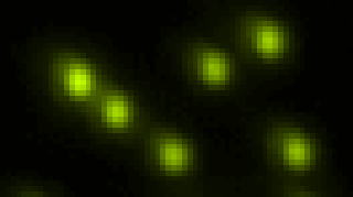
 Theoretical PSF
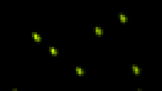
 Experimental PSF
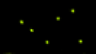

More information
See Deconvolving Beads.