
Because a Refractive Index Mismatch distorts the PSF, that has a straightforward effect for example in the image of recorded beads. See Imaging Simulations below for the images of a 0.1 µm (diameter) fluorescent bead when recorded in a Confocal Microscope a depth zero (no Spherical Aberration) and at depth 20 µm inside the sample, having a Refractive Index Mismatch 1.515 - 1.47 and with a Signal To Noise Ratio of 15.
The images are 2 µm wide.
Top view Maximum Intensity Projection
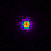
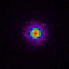
(False colors). The differences in the XY planes are apparently not very significative, but the aberrated image has noticeable intensity at a larger extent around the center.
XZ slice
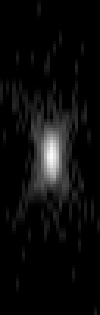
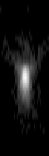
(Grey-scale with Gamma Correction). The image is clearly elongated along Z.
Microscopic Parameters in this Imaging Simulation: x-y Sample Size (nm) </td><td> 63.5 z Sample Size (nm) </td><td> 200 Microscope Type </td><td> Confocal Microscope Numerical Aperture </td><td> 1.3 Pinhole Radius (nm) </td><td> 307 Lens Refractive Index </td><td> 1.515 (Oil) Medium Refractive Index </td><td> 1.47 Excitation Wavelength (nm) </td><td> 568 Emission Wavelength (nm) </td><td> 575 Excitation Photons </td><td> 1